Sept. 20, 2022

Facet Joint Syndrome / Arthritis


Facet Joint Syndrome


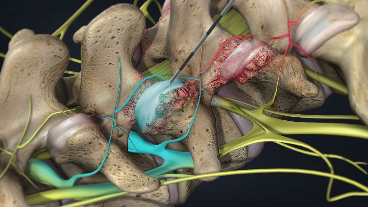
Anatomy of the facet joints

Facet Joint Syndrome is a condition in which arthritic change and inflammation occur, and the nerves to the facet joints convey severe and diffuse pain.

the facet joints are the connections between the bones of the spine. They allow the spine to bend and twist and they prevent excessive forward slipping or twisting. Like knee joints, they contain cartilage for smooth movement where two bones meet. The joints are lined with synovium and have lubricating joint fluid.

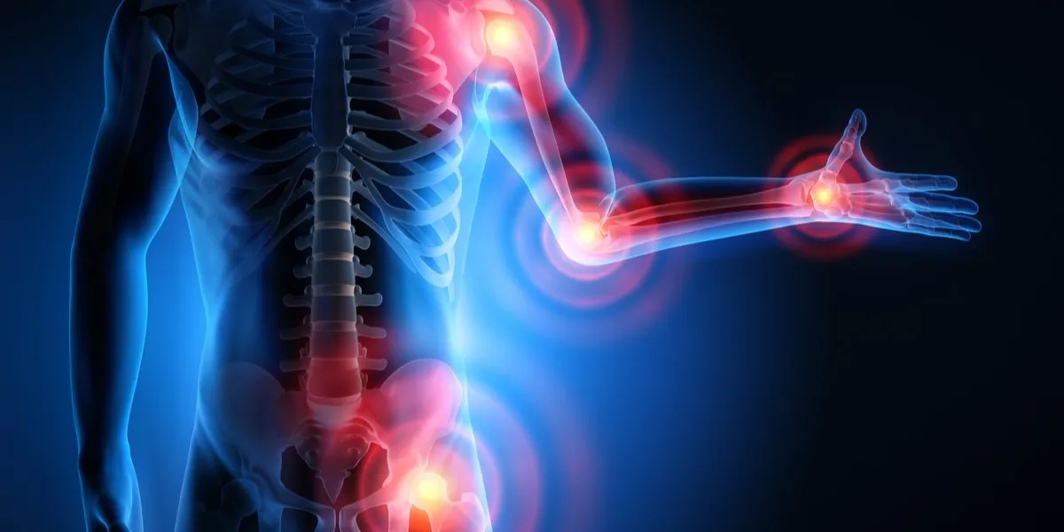
When these joints become inflamed due to injury or arthritis, they cause pain. If the affected joint is in the neck, it may lead to headaches and difficulty moving the head. If it is in the back, it may cause pain in the lower back, buttocks, or thighs.

The spine comprises movable bones called vertebrae, each of which functions as a three-joint complex with a large disc in the front and two facet joints in the back. This robust design keeps the bones linked together while allowing the spine to bend and twist. Facet joints are synovial joints that are lined with cartilage, lubricated with synovial fluid, and covered by a joint capsule. Healthy facet joints allow for smooth gliding and sliding movements in the back while preventing over-twisting.


Facet Arthritis

What is facet joint syndrome?

Pain that comes from one or more facet joints is called facet joint syndrome or facet arthropathy. Degenerative changes in the spine may cause body weight to shift unevenly to the facet joints. This extra burden causes wear and tear on the joint and change it over time. The joint capsule thins, smooth cartilage breaks down and becomes irregular like a cobblestone street, and bone spurs may form. Similar to arthritis of the knee joint, these changes make it difficult for the joint to move fluidly and it reacts by becoming inflamed and irritated. The irritated joint sends pain signals to the brain via small nerves in the capsule called medial branch sensory nerves. In turn, the muscles in the area can stiffen and spasm.

In short, injury and degeneration cause the natural movement of these joints to break down, and the resulting inflammation, nerve irritation, and uneven pressure can cause intense pain and a significant decrease in your quality of life.

Common Signs and Symptoms of Facet Joint Pain

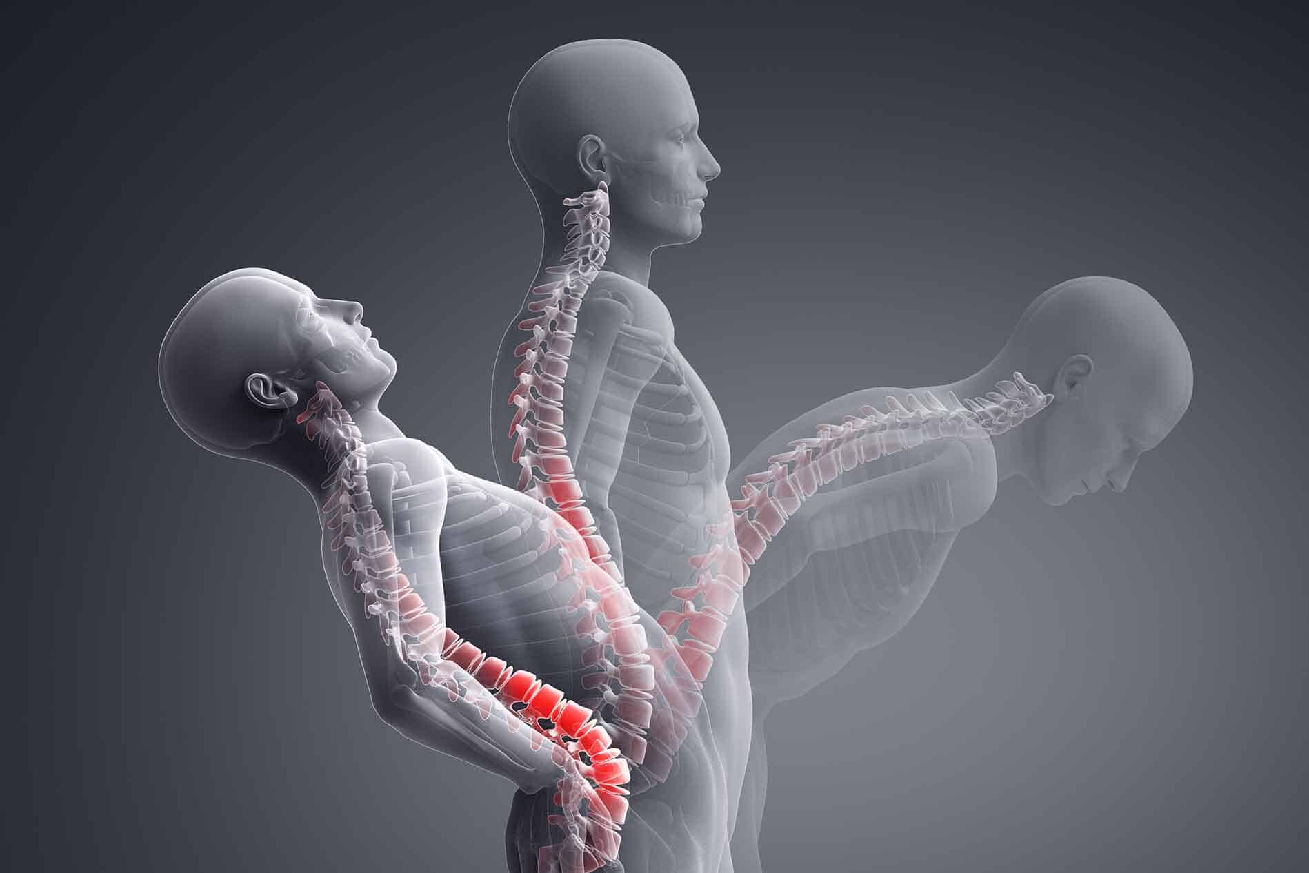
Facet joint pain can occur gradually, in the case of degeneration, or suddenly when caused by injury. The most common symptoms of Facet Joint Syndrome are:

  • Difficulty twisting and bending the spine: These kinds of movements specifically exasperate the pain, which can make the activities of daily living difficult for you to perform.
  • Pain and inflammation: The most commonly affected areas for pain and inflammation are your lower back and neck.
  • Numbness, tingling, and muscle weakness: These occur when the edges of one of your facet joints rub on a nerve, which can also be very painful.
  • Bone spurs: When your facet joints don’t line up correctly, your bones certainly will meet where they shouldn’t and rub against each other. Unsurprisingly, this also becomes very painful.
  • Tenderness of certain areas of the spine: Occurs in response to inflammation in the nerves and muscles surrounding your facet joints.

These symptoms affect different parts of your body depending on which spinal nerves have been irritated.

  • Neck pain and stiffness, such as might force you to turn your entire body to look at something, may indicate Cervical Facet Joint Syndrome.
  • Pain near the middle of your back may indicate Thoracic Facet Joint Syndrome. Joint hypertrophy is less common in the thoracic spine due to the natural rigidity of the vertebrae.
  • Low back pain and stiffness, such as might make it difficult to stand up from a chair or straighten your back, may indicate Lumbar Facet Joint Syndrome.
  • Spinal stenosis can also occur in these areas, which narrows the spaces within your spine. This puts pressure on the nerves that travel through your spine to the rest of your body.

What are the causes?

As we get older, the cartilage in our joints wears down. An injury, repetitive movements, obesity, poor posture, and other spine conditions that change the way the facet joints align and move can cause pain.

Changes in the facet joints can begin with the deterioration of a vertebral disc. As the load of the body weight shifts to the facet joint, the cartilage breaks down, the joint space narrows, and the bones rub together.

Who is affected?

Facet joint syndrome occurs in both men and women. It is most common between the ages of 40 and 70 and in those prone to arthritis. It also may develop in people who’ve had a spine injury.

How does your osteopath diagnose a Facet Joint Injury?

The most accurate diagnosis of a facet joint injury is by palpation/touch. Osteopaths are trained to examine the spine, and they develop a remarkable sense of touch.

You may be asked to stand or move in different positions and point to where you feel pain. The osteopath may manipulate your joints or feel for tenderness over the spine.

Imaging studies, such as X-rays, CTs, or MRIs, may be ordered to help diagnose and check for other spine and hip-related problems, but some of the diagnostic tools you might expect a doctor to use are not needed in these cases. X-rays, MRI, and CT scans are useful to identify arthritic changes and fractures but are unable to detect a locked facet joint.

Facet Joint Injections can also be used to help identify facet joint pain. The facet joint is injected with a local anesthetic and corticosteroid medication. The injection is given using X-ray fluoroscopy to ensure accurate needle placement in the facet joint. Your pain level is evaluated before and 20-30 minutes after injection, and monitored over the next week. Facet joint involvement is confirmed if your pain level decreases by more than 75%. If your pain level does not change after the injection, it is unlikely that the facet joint is the cause of your pain.

Conservative Treatments for Facet Joint Pain

Pain from Facet Joint Syndrome can be treated conservatively with postural correction, physical therapy, massage, and chiropractic adjustment. These treatments can reestablish normal motion and increase the strength of the surrounding muscles to help support your spine. Conservative treatments are usually combined with anti-inflammatory medications and possibly muscle relaxers to diminish your acute pain while your body heals.

Most conservative treatments for facet syndrome involve postural correction, soft tissue massage, and manipulation of the affected areas.

Osteopathic clinicians and chiropractors are adept at restoring restricted and painful facet joints to move and can reestablish normal motion. Treatments are usually combined with a course of anti-inflammatory medications to decrease inflammation. Muscle relaxers may be used to decrease local muscle spasms.

In some cases, the pain may be so severe or the condition so advanced that conservative therapies are either too painful or otherwise ineffective. In these cases, a patient may be recommended for interventional pain therapy.

Minimally-Invasive Pain Therapy for Facet Joint Syndrome

If conservative treatments are ineffective or too painful, you may be recommended for one or more minimally-invasive surgical procedures. These procedures can be performed on an outpatient basis, and are done safely using fluoroscopic X-ray guidance. Interventional pain management therapies can offer rapid pain relief and help you resume normal activity.

Minimally-invasive treatment options may include:

  • Facet Joint Injections: This procedure injects a corticosteroid into the affected joint which reduces pain and inflammation. These injections may also be used as part of the diagnostic process for additional treatments. Facet joint pain, but there is a limit on how frequently they can be administered.
  • Facet Joint Block: This procedure is similar to a facet joint injection, but instead injects a local anesthetic into each side of the vertebrae. This will temporarily numb the pain and confirm the source of the injury.
  • Radiofrequency Ablation: By destroying (ablating) the nerve endings surrounding the affected facet joint, pain signals can be blocked which will provide you with relief. This procedure often begins with a median nerve block to confirm the affected nerves for ablation.
Lumbar Spine Facet Joint Dysfunction And Pain

How We Can Help

If you have questions about how York rehab clinic can help you get back to a life free from the pain of Facet Joint Syndrome. Our osteopaths and chiropractors specialize in conservative and none-invasive treatment plans for back, neck, and joint pain.

To book an appointment for Osteopathy, Chiropractic, and massage therapy in Richmond Hill, contact our professional team at York Rehab Clinic today at (416) 350-1940.

Arthritis – Symptoms and Treatments

Arthritis – Symptoms and Treatments

Introduction: Arthritis is a common and often painful condition that affects millions of individuals worldwide. It’s not a single disease but rather a group of more than 100 different types of joint-related conditions. In this comprehensive article, we will delve into Arthritis, exploring its causes, symptoms, effective treatment options, including the crucial roles of physiotherapy and osteopathy. We’ll also touch on related diseases and provide valuable insights for managing this condition.     Understanding Arthritis: Arthritis is a complex and multifaceted health challenge that primarily affects the joints. These vital connectors in our bodies allow for movement, but when afflicted by arthritis, they can become sources of chronic pain and discomfort. It’s essential to recognize that arthritis doesn’t discriminate – it can affect individuals of all ages, genders, and backgrounds.     The Complexity of Arthritis: The term Arthritis encompasses a wide array of conditions, each with its unique characteristics. Among the most common forms are osteoarthritis and rheumatoid arthritis. Osteoarthritis, often associated with the wear and tear of joints over time, is a leading cause of pain and mobility issues in older adults. Rheumatoid arthritis, on the other hand, is an autoimmune disorder where the body’s immune system mistakenly attacks the joints, leading to inflammation and pain.     Causes of Arthritis: Arthritis is a multifaceted condition with various underlying causes, and understanding these triggers is essential for effective management. Here, we delve deeper into the key factors contributing to arthritis: Age and Wear and Tear: Osteoarthritis, the most prevalent form of arthritis, often develops with age as joint tissues naturally degenerate over time. Repetitive use and wear and tear on joints can accelerate this process. Genetics: Family history plays a significant role in some forms of arthritis. If close relatives have arthritis, there may be a genetic predisposition that increases the risk of developing the condition. Autoimmune Responses: Rheumatoid arthritis and other autoimmune forms of arthritis occur when the body’s immune system mistakenly attacks healthy joint tissues. These conditions are characterized by chronic inflammation and joint damage. Infections: In rare cases, infections can trigger reactive arthritis. Bacterial or viral infections elsewhere in the body can lead to joint inflammation. Metabolic Abnormalities: Conditions like gout result from metabolic abnormalities, particularly elevated levels of uric acid in the bloodstream. This excess uric acid can crystallize in joints, causing intense pain and inflammation.     Symptoms of Arthritis: Arthritis presents a wide spectrum of symptoms that can significantly impact an individual’s daily life. Recognizing these signs is crucial for early diagnosis and effective management. Here, we outline the common symptoms associated with arthritis: Joint Pain: Persistent and often severe joint pain is a hallmark of arthritis. The pain can range from a dull ache to sharp, shooting sensations and may be more pronounced during movement. Joint Swelling: Inflammation of the joints leads to swelling, which can be visibly apparent and cause joint deformities over time. Stiffness: Arthritis-related stiffness can make it challenging to initiate movement, particularly after periods of inactivity, such as waking up in the morning. Limited Range of Motion: Reduced flexibility and range of motion in affected joints can hinder daily activities like bending, reaching, or walking. Fatigue: Many individuals with arthritis experience fatigue, often attributed to the body’s constant battle against inflammation. Weakness: Muscles around affected joints may weaken due to reduced use, leading to further functional limitations. Numbness and Tingling: In some cases, nerve compression or irritation related to arthritis can result in sensations of numbness or tingling in the hands or feet. Fever and Weight Loss: Systemic symptoms like fever and unexplained weight loss can accompany certain forms of inflammatory arthritis. Joint Warmth and Redness: Inflammation can cause affected joints to become warm to the touch and appear red or flushed. It’s crucial to remember that the severity and combination of these symptoms can vary widely among individuals and arthritis types. Early consultation with healthcare professionals and a tailored treatment plan, which may include physiotherapy and osteopathy, can significantly improve one’s quality of life when living with arthritis.     Types of Arthritis: Arthritis isn’t a single, uniform condition; it encompasses a diverse group of disorders, each with its unique characteristics and underlying causes. Understanding the various types of arthritis is essential for accurate diagnosis and effective management. Here, we explore some of the most prevalent types: Osteoarthritis (OA): Osteoarthritis is the most common form of arthritis, typically associated with the natural aging process. It occurs as the protective cartilage that cushions the ends of bones deteriorates over time. OA commonly affects weight-bearing joints like the knees, hips, and spine, causing pain, stiffness, and reduced mobility. Rheumatoid Arthritis (RA): Rheumatoid arthritis is an autoimmune disorder in which the body’s immune system mistakenly attacks the synovium—the lining of the membranes that surround the joints. This leads to chronic inflammation, joint damage, and pain. RA often affects joints on both sides of the body, such as wrists, knees, and fingers. Psoriatic Arthritis: Psoriatic arthritis is a type of inflammatory arthritis that occurs in individuals with psoriasis, a skin condition characterized by red, scaly patches. It can affect any joint and may involve the fingers and toes, causing swelling, pain, and stiffness. Ankylosing Spondylitis: Ankylosing spondylitis primarily affects the spine and sacroiliac joints, leading to chronic inflammation and pain. Over time, it can cause the vertebrae to fuse, resulting in reduced mobility. This condition often starts in early adulthood and disproportionately affects men. Gout: Gout is caused by the buildup of uric acid crystals in the joints, resulting in sudden and severe joint pain, often in the big toe. Dietary factors and genetics play a role in its development. Gout flares can be managed with medications and lifestyle changes. Juvenile Arthritis: Juvenile arthritis refers to a group of autoimmune disorders that affect children under the age of 16. The most common form is juvenile idiopathic arthritis (JIA), which causes joint inflammation, pain, and stiffness. Early diagnosis and treatment are crucial to prevent long-term joint damage. Lupus (Systemic Lupus Erythematosus): While primarily known as a systemic autoimmune disease, lupus can also manifest as a form of

Hallux Valgus (Bunions)

Hallux Valgus (Bunions)

Overview Hallux valgus, commonly known as a bunion, is a complex foot deformity that occurs when the bones of the big toe move out of alignment. This misalignment pushes the big toe toward the smaller toes, causing a bony bump at the base of the toe, which can become red, swollen, and painful. The condition can make daily activities uncomfortable and may worsen over time if left untreated. Bunions typically develop due to pressure on the big toe, often from wearing tight, narrow shoes, though they can also be caused by foot deformities, arthritis, or the natural shape of the foot. The deformity is easily diagnosable through a physical exam, with imaging used to assess any potential damage to the joint. Initial treatment for hallux valgus focuses on non-surgical options, including wearing wider shoes, using orthotics, and applying night splints to relieve pressure and correct the alignment. Addressing symptoms such as pain and stiffness is crucial to improving quality of life. In some cases, smaller bunions, known as bunionettes, can also develop on the joint of the little toe. I am text block. Click edit button to change this text. Lorem ipsum dolor sit amet, consectetur adipiscing elit. Ut elit tellus, luctus nec ullamcorper mattis, pulvinar dapibus leo.   Types and Prevalence of Bunions Bunions, particularly on the big toe, are common foot deformities that result from pressure on the toe joint. However, they can also occur on other toes or for different reasons. There are several types of bunions: Congenital Bunions (Congenital Hallux Valgus): These bunions are present at birth, resulting from foot structure abnormalities. Juvenile or Adolescent Hallux Valgus: This type affects individuals younger than 18 and develops due to inherited foot structure or other factors. Tailor’s Bunion (Bunionette): A bunionette forms at the base of the little (pinky) toe, often caused by tight shoes or activities that put pressure on the small toe.   How Common Are Bunions? Bunions are quite prevalent , Hallux valgus deformity, the most common type, affects up to 30% of adult females, particularly those who wear tight shoes or high heels. Women are found to have this deformity twice as often as men, especially in populations that wear shoes compared to those who go barefoot.     Symptoms and Causes of Bunions Symptoms: Bunions are often characterized by a noticeable bony bump at the base of the big toe. In addition to the visible bump, other common symptoms include: – Swelling, redness, or soreness around the big toe joint. – Pain or stiffness in the big toe, which can worsen when wearing shoes. – Limited movement of the big toe, potentially accompanied by a burning sensation. – Corns or calluses that form where the toes rub against each other. – Numbness around the big toe. – In severe cases, hammertoes (tight, painful toe tendons) may also develop. Causes: While the exact cause of bunions is not definitively known, several contributing factors are thought to play a role. These include: – Inherited Foot Type: Genetic factors may predispose some individuals to develop bunions. – Foot Stress or Injuries: Repeated stress or trauma to the foot can push the joint out of alignment. – Congenital Deformities: Some individuals are born with foot deformities that make bunions more likely. – Shoes: Wearing narrow, tight, or high-heeled shoes can exacerbate the problem, though experts disagree on whether they directly cause bunions. – Health Conditions: Inflammatory conditions like rheumatoid arthritis or lupus can contribute to bunion formation. – Foot Mechanics: Abnormal walking patterns or standing for prolonged periods can increase pressure on the big toe joint, leading to bunions over time.     Related Conditions: Bunions can often be associated with or lead to other foot problems, including: Bursitis: Inflammation of the fluid-filled sac (bursa) near the joint. Hammer toe: A condition where the middle joint of a toe becomes bent, resembling a hammer. Metatarsalgia: Pain and inflammation in the ball of the foot. Toe Overlap: The big toe may overlap or underlap adjacent toes.     Treatment Options: Effective treatment for bunions focuses on alleviating pain, slowing the progression of the condition, and improving foot function. Treatment options include: Footwear: Choosing comfortable, well-fitting shoes with a wide toe box can reduce pressure on the bunion and prevent worsening of the condition. Orthotics: Custom-made or over-the-counter shoe inserts can help redistribute pressure and provide arch support, reducing discomfort. Medications: Over-the-counter pain relievers or anti-inflammatory medications may provide temporary relief from pain and inflammation. Icing: Applying ice to the bunion can help reduce inflammation and alleviate pain. Padding and Taping: Using cushions or tape can protect the bunion from friction and pressure. Physical Therapy: Physiotherapy and osteopathy techniques can enhance foot and joint mobility, aiding in the management of bunion symptoms.   Home Remedies for Bunion Pain Relief: In addition to medical treatment, individuals with bunions can try home remedies to alleviate pain and discomfort: Warm Soaks: Soaking your feet in warm water can help reduce pain and swelling. Toe Spacers: Using toe spacers or cushions can help realign the toes and reduce pressure on the bunion. Proper Footwear: Wearing shoes with ample room for the toes and good arch support can alleviate discomfort. Bunion Pads: Applying bunion pads or cushions can reduce friction and irritation. Gentle Exercises: Practicing exercises to strengthen the muscles in the foot can help with alignment. Weight Management: Maintaining a healthy weight can reduce pressure on the feet, potentially slowing bunion progression.     Conclusion: Bunions are a common foot issue that can cause discomfort and affect daily activities. Early intervention, proper footwear, and non-invasive treatments like physiotherapy and osteopathy can help manage symptoms effectively. For severe cases, surgical options may provide lasting relief. If you suspect you have bunions or are experiencing foot pain, consult a healthcare provider or podiatrist for an accurate diagnosis and personalized treatment plan. Preventive measures, such as wearing suitable footwear and paying attention to foot health, can also reduce the risk of developing bunions.

Muscular Dystrophy: Causes andTreatments

Muscular Dystrophy: Causes andTreatments

Introduction Muscular dystrophy is a group of genetic disorders characterized by progressive muscle degeneration and weakness. The condition stems from mutations in the genes responsible for the production of specific muscle proteins, leading to the gradual deterioration of muscle mass and function over time. While various types of muscular dystrophy exist, the most common forms typically manifest during childhood, predominantly in boys. However, certain variations might not become evident until adulthood.     Symptoms The primary indicator of muscular dystrophy is the gradual weakening of muscles. Different signs and symptoms arise at varying ages and in diverse muscle groups, depending on the specific type of muscular dystrophy. Duchenne Type Muscular Dystrophy: Frequent falls Difficulty in rising from a lying or sitting position Challenges with running and jumping Waddling gait Walking on the toes Enlarged calf muscles Muscle pain and stiffness Learning disabilities Delayed growth Becker Muscular Dystrophy: Symptoms resembling Duchenne Muscular Dystrophy, albeit milder and slower in progression.     Causes Muscular dystrophy originates from genetic mutations that impair the production of essential muscle proteins, resulting in the degeneration and weakening of muscle tissues. These mutations can be inherited or arise spontaneously, contributing to the development of specific forms of the disease. Understanding the underlying genetic mechanisms responsible for each type of muscular dystrophy is crucial for accurate diagnosis and targeted treatment approaches.     Types of muscular dystrophy Duchenne Type Muscular Dystrophy: Duchenne muscular dystrophy is the most prevalent form of the condition, primarily affecting boys. This type typically manifests in early childhood and is characterized by progressive muscle weakness and wasting, leading to difficulties with mobility and motor functions. Common symptoms include frequent falls, trouble rising from a sitting or lying position, a waddling gait, and enlarged calf muscles. As the disease advances, it can also result in learning disabilities and delayed growth, significantly impacting the overall quality of life for affected individuals. Becker Muscular Dystrophy: Becker muscular dystrophy shares similarities with Duchenne muscular dystrophy, although the progression is generally slower and the symptoms milder. This type of muscular dystrophy often presents in the teenage years or even in adulthood, with manifestations such as muscle weakness, difficulties with movement, and potential complications related to mobility and muscle function. While it bears resemblance to Duchenne muscular dystrophy, the milder progression often allows for a more extended period of preserved mobility and functionality. Myotonic Dystrophy: Myotonic dystrophy is characterized by the inability of muscles to relax following contractions. This type typically affects facial and neck muscles initially, resulting in distinct physical features such as a long, thin face, drooping eyelids, and a swan-like neck. Apart from the facial and neck muscles, myotonic dystrophy can also involve other muscle groups, leading to a range of symptoms including muscle weakness, myotonia (prolonged muscle contractions), and potential complications affecting mobility and muscle function. Facioscapulohumeral Muscular Dystrophy (FSHD): Facioscapulohumeral muscular dystrophy primarily affects the face, shoulder, and hip muscles, often leading to the appearance of winged shoulder blades when the arms are raised. Onset can occur during the teenage years or later in life, presenting with symptoms such as muscle weakness, difficulties with movement, and progressive impairment of mobility. The variable age of onset and differing patterns of muscle involvement contribute to the diverse clinical presentation and progression of FSHD. Congenital Muscular Dystrophy: Congenital muscular dystrophy manifests early in life, either at birth or before the age of two, and can result in a range of outcomes depending on the specific form of the condition. Some variations progress gradually, leading to mild disabilities, while others can rapidly cause severe impairments. The symptoms may include muscle weakness, developmental delays, joint deformities, and potential complications related to mobility and motor functions, necessitating comprehensive management and support from an early age. Limb-Girdle Muscular Dystrophy: Limb-girdle muscular dystrophy affects the muscles around the hips and shoulders, resulting in difficulties with lifting the front part of the foot and frequent tripping. The onset of this type of muscular dystrophy typically occurs during childhood or the teenage years, leading to progressive muscle weakness, mobility challenges, and potential complications related to muscle function and overall physical activity. The diverse subtypes of limb-girdle muscular dystrophy contribute to the variability in the presentation and progression of the condition, necessitating tailored treatment approaches and comprehensive support for affected individuals.     Clinic Services for Muscular Dystrophy: At York Rehab Clinic, our comprehensive range of services is tailored to effectively address the management of muscular dystrophy. With a focus on providing holistic and personalized care, our clinic combines various treatment modalities to support individuals with muscular dystrophy in their journey toward improved mobility and enhanced quality of life. Physiotherapy and Osteopathy: These services play a pivotal role in maintaining muscle strength, flexibility, and overall physical function. Through targeted exercises, manual therapy, and specialized techniques, our experienced physiotherapists and osteopaths work closely with patients to enhance muscle tone, improve joint mobility, and alleviate discomfort associated with muscular dystrophy. Shockwave Therapy: This innovative treatment option offered at York Rehab Clinic aids in promoting tissue regeneration and reducing pain through the application of focused shockwave energy. By stimulating the body’s natural healing mechanisms, shockwave therapy can effectively address muscle soreness and improve overall muscle function in individuals with muscular dystrophy. Knee and Spinal Decompression Therapy: Our clinic provides specialized decompression therapies, including knee and spinal decompression, to alleviate pressure on affected joints and spinal structures. By creating a controlled and gentle traction force, these therapies contribute to relieving pain and improving mobility, particularly in areas commonly affected by muscular dystrophy. Acupuncture: As an integral component of our treatment approach, acupuncture serves as a valuable complementary therapy in managing muscular dystrophy. By targeting specific acupoints, this technique can help reduce muscle tension, alleviate pain, and enhance overall well-being, promoting a comprehensive approach to managing the symptoms associated with the condition. PRP (Platelet-Rich Plasma) Therapy: At York Rehab Clinic, PRP therapy is utilized as an innovative regenerative treatment option to support muscle repair and enhance tissue healing. By harnessing the body’s

Gluteal Tendinopathy: Causes -Treatments

Gluteal Tendinopathy: Causes -Treatments

Introduction: The Gluteal tendons are robust bands of tissue that connect the deep buttock muscles (gluteus medius and minimus) to the hip bone at a site known as the greater trochanter. Gluteal Tendinopathy is sometimes referred to as Greater Trochanteric Pain Syndrome (GTPS). These tendons play a crucial role in transferring the force generated by your gluteal muscles to the thigh, enabling a range of movements such as lifting your leg out to the side and maintaining pelvic stability during activities like walking, running, squatting, climbing stairs, and rising from a chair.     What is Gluteal Tendinopathy? Gluteal Tendinopathy refers to the irritation or damage to the tendons in the gluteal region, leading to pain and limited mobility. This condition is often associated with overuse, muscle imbalances, or traumatic injuries that affect the integrity of the gluteal tendons.     Symptoms: Persistent pain and tenderness in the lateral hip and buttock area, often worsening during activities such as walking, climbing stairs, or prolonged sitting. Discomfort or pain that can radiate down the outside of the thigh. Stiffness and limited range of motion in the affected hip joint, leading to difficulties in performing daily activities. Weakening of the gluteal muscles, resulting in compromised hip strength and stability. Changes in gait patterns characterized by limping or favoring one side over the other. In some cases, individuals may experience inflammation and swelling in the affected hip area, contributing to heightened discomfort and restricted movement.     Causes: Overuse or repetitive stress on the gluteal tendons, often seen in athletes or individuals engaging in activities that involve frequent hip movements, such as running or cycling. Muscle imbalances or weakness in the hip and core muscles, leading to altered biomechanics and increased strain on the gluteal tendons. Direct trauma or injury to the hip area, resulting from falls, accidents, or sudden impacts during sports or physical activities. Degeneration of the tendons due to aging, underlying medical conditions, or a history of chronic inflammation in the hip region.     Effective Treatments for Gluteal Tendinopathy: At York Rehab Clinic, we provide a range of specialized services aimed at effectively managing gluteal Tendinopathy. Our comprehensive treatment approach includes: Physiotherapy: Our skilled physiotherapists utilize tailored exercise programs and manual therapy techniques to improve muscle strength, flexibility, and joint stability in the gluteal region. Osteopathy: With a focus on holistic musculoskeletal care, our osteopathic treatments aim to alleviate pain, improve tissue mobility, and restore balance in the hip and buttock area, promoting comprehensive functional recovery. Shockwave Therapy: This advanced treatment modality stimulates tissue regeneration, reduces pain, and accelerates the healing process, providing a non-invasive option for managing gluteal Tendinopathy effectively. Acupuncture: As a complementary therapy, acupuncture can help relieve muscle tension, reduce pain, and enhance overall well-being, serving as a valuable adjunct to traditional treatment approaches for gluteal tendinopathy.       What’s the difference between gluteal tendinopathy, gluteal tendinitis and trochanteric bursitis? While gluteal Tendinopathy, gluteal tendinitis, and Trochanteric bursitis share some common symptoms and affect the hip and buttock region, each condition has distinct characteristics and underlying causes. Gluteal Tendinopathy: Gluteal Tendinopathy refers to a chronic condition involving the degeneration of the gluteal tendons, which connect the gluteal muscles to the hip bone. This condition typically develops due to repetitive stress or overuse of the tendons. It can cause persistent pain and stiffness in the hip region, leading to mobility issues and discomfort during activities such as walking, climbing stairs, or sitting for extended periods. Gluteal Tendinopathy is often characterized by a gradual onset of symptoms and may progress if left untreated. Gluteal Tendinitis: Gluteal tendinitis specifically refers to the inflammation of the gluteal tendons. It commonly occurs as a result of repeated stress or injury to the tendons, leading to localized pain, tenderness, and swelling in the hip area. Individuals with gluteal tendinitis often experience discomfort during movements that involve the gluteal muscles, such as running, squatting, or prolonged sitting. The condition can affect mobility and may become more aggravated with certain activities or positions. Trochanteric Bursitis: Trochanteric bursitis is characterized by the inflammation of the bursa, a small, fluid-filled sac located near the outer point of the hip, known as the greater trochanter. This condition often arises from repetitive friction or stress on the bursa, leading to pain and tenderness in the hip region, particularly during physical activities that involve side-to-side movements or prolonged pressure on the hips. Trochanteric bursitis can impact daily activities and may cause discomfort during sleep when lying on the affected side. Understanding the distinctions between gluteal Tendinopathy, gluteal tendinitis, and Trochanteric bursitis is essential for accurate diagnosis and the implementation of effective treatment strategies. At York Rehab Clinic, our specialized team provides personalized care to address these conditions comprehensively. For further information or to schedule a consultation, please contact us at 416-350-1940 or visit our website at www.yorkrehabclinic.ca. You can find us conveniently located at 10825 Yonge St Unit 5, Richmond Hill, ON L4C 3E3.


Shoulder Impingement (Rotator Cuff Tendinitis)


Shoulder Impingement (Rotator Cuff Tendinitis)

Overview What is Shoulder Impingement? Shoulder impingement occurs when the rotator cuff tendons become compressed by the bones of the shoulder joint, particularly the upper part of the shoulder blade (scapula). This condition, often referred to a shoulder impingement syndrome, leads to pain and discomfort, especially during arm movement.Shoulder impingement is common among athletes, manual laborers, and people who engage in repetitive overhead activities. The rotator cuff consists of four muscles that connect the shoulder blade to the upper arm bone (humerus), allowing you to lift and rotate your arm. These muscles are crucial for overhead movements and maintaining shoulder stability. Because the rotator cuff sits in a narrow space between the upper arm bone and the shoulder blade, it can easily become irritated or “impinged” between these bones. This pinching leads to inflammation, pain, and reduced mobility in the shoulder. If you are experiencing shoulder pain or difficulty moving your shoulder without discomfort, it’s important to visit a healthcare provider for proper evaluation and treatment.     Types of Shoulder Impingement Syndrome Healthcare providers may categorize shoulder impingement into more specific conditions, including: Rotator Cuff Tendinitis Rotator cuff tendinitis is an inflammation or irritation of the tendons in the rotator cuff. The rotator cuff consists of four tendons that help move and stabilize the shoulder joint. When these tendons become inflamed—due to overuse, injury, or strain—it leads to swelling, which causes pain and limits shoulder movement. This swelling also reduces the space within the shoulder joint, resulting in impingement of the tendons. Shoulder Bursitis Shoulder bursitis refers to the inflammation of a bursa, which is a small fluid-filled sac that cushions joints. In the shoulder, a bursa located between the rotator cuff and the acromion (the top part of the shoulder blade) helps reduce friction during movement. When this bursa becomes inflamed, the swelling can cause discomfort and contribute to the impingement of the shoulder tendons, leading to symptoms like pain and limited mobility. Acromion Deformity The acromion is a part of the shoulder blade that forms the roof of the shoulder joint. In a healthy shoulder, the acromion is usually flat. However, some individuals may have a curved or hooked acromion, either due to genetics or as a result of bone changes with age. These deformities can lead to increased pressure on the rotator cuff, causing pinching and impingement. Additionally, the development of bone spurs in this area can further narrow the space and aggravate the impingement.     Symptoms and Causes Symptoms of Shoulder Impingement Shoulder impingement typically manifests as pain that primarily originates in the front of the shoulder. Individuals may experience: Tenderness: The affected area may feel sensitive when touched. Radiating Pain: Discomfort can extend from the front of the shoulder down the arm. Nocturnal Pain: Pain may intensify at night, disrupting sleep. Aggravated Symptoms: Certain movements can exacerbate pain, including: Raising the arm overhead. Lifting or lowering objects. Reaching for items. Lying on the side of the affected shoulder. Reaching behind the back, such as when accessing a pocket or unzipping a bag. In addition to pain, some people may notice other signs, such as stiffness in the shoulder, swelling, or a sensation of popping or cracking during movement.   Causes of Shoulder Impingement Shoulder impingement is primarily an overuse injury, often resulting from repetitive activities that place excessive strain on the shoulder joint and rotator cuff. This can happen due to: Repetitive Motions: Engaging in activities that involve overhead movements, like certain sports or labor-intensive jobs, can lead to irritation and inflammation of the shoulder tendons. Injury or Trauma: Sudden impacts from falls, accidents, or sports injuries can trigger impingement. In some cases, shoulder impingement develops without a clear cause, referred to as idiopathic impingement.     Risk Factors for Shoulder Impingement While anyone can develop shoulder impingement, certain groups are at higher risk. Individuals involved in athletics or physically demanding occupations are particularly susceptible. Common sports associated with shoulder impingement include: Swimming Baseball Volleyball Tennis Occupations that require repetitive shoulder movements, such as: Construction painting Window washing Hanging wallpaper or drywall can also increase the likelihood of developing this condition. Symptoms often emerge gradually, worsening over time as the shoulder sustains repeated strain.     Management and Treatment Conservative Treatments Rest: Avoid activities that worsen the symptoms, particularly repetitive overhead motions. Ice application: Applying ice to the shoulder can help reduce inflammation and pain. Non-steroidal anti-inflammatory drugs (NSAIDs): Medications like ibuprofen can be used to manage pain and inflammation. Stretching and strengthening exercises: Physical therapy can focus on stretching tight muscles and strengthening the shoulder to improve function and reduce impingement. Medical Treatments Corticosteroid injections: Steroid injections may be recommended to reduce inflammation and provide pain relief. Laser Therapy: Laser therapy can promote tissue healing by reducing inflammation and improving circulation to the affected area. Surgery: In severe cases, surgery might be needed to remove bone spurs or other structures that are contributing to impingement.     Prevention Preventing shoulder impingement involves maintaining good shoulder mechanics and posture: Strengthening exercises: Focus on strengthening the rotator cuff and surrounding muscles to support shoulder stability. Proper posture: Keeping the shoulders back and avoiding slouching helps reduce strain on the rotator cuff. Avoid repetitive overhead activities: Whenever possible, minimize activities that put excessive strain on the shoulder joint. Warm-up exercises: Prior to any physical activity, warm up your shoulder with light stretches to prevent injury.     Care at York Rehab Clinic: Laser Therapy and Physiotherapy At York Rehab Clinic, we offer comprehensive treatment options to manage shoulder impingement and promote healing. Laser Therapy Our advanced laser therapy uses targeted light energy to reduce inflammation, promote tissue regeneration, and accelerate healing in the shoulder tendons. This non-invasive therapy is effective in relieving pain and improving mobility in patients with shoulder impingement. Physiotherapy Our specialized physiotherapy programs focus on restoring shoulder strength and flexibility. We use tailored exercises to improve muscle balance, correct posture, and alleviate the pain caused by rotator cuff

Achondroplasia – Symptoms, Causes

Achondroplasia – Symptoms, Causes

what is Achondroplasia? Achondroplasia is a genetic disorder causing short-limb dwarfism due to improper cartilage development in the arms and legs. It results from a mutation in the FGFR3 gene, which disrupts normal bone growth. Adults with achondroplasia typically reach heights of 42 to 56 inches. This condition can also lead to neurological issues in 20 to 50 percent of affected children. Managing achondroplasia involves genetic counseling and medical care to address related health concerns.     What is the difference between achondroplasia and skeletal dysplasia (dwarfism)? Achondroplasia is the most common form of skeletal dysplasia, which is the umbrella term used to identify hundreds of conditions that affect the growth of bones and cartilage. Achondroplasia specifically targets bone growth in your arms and legs.     Is achondroplasia hereditary? Most cases of achondroplasia are not inherited. Anyone can be affected by achondroplasia. Around 80% of individuals with achondroplasia have parents of normal height and are born with a new gene alteration (de novo mutation). It is rare that these parents will have another child with achondroplasia. Only one parent needs to pass down the gene for a child to be born with achondroplasia (autosomal dominant). There is a 50% chance of a person with achondroplasia and a partner who does not have achondroplasia having a child with the disorder. If both parents have achondroplasia, there is a 25% chance that the child will be born with homozygous achondroplasia, which leads to stillbirth or death shortly after birth.     Symptoms Achondroplasia, a genetic disorder, manifests through a variety of physical characteristics and developmental delays. Common symptoms include: Shortened Limbs: Upper arms and thighs are more affected than forearms and lower legs. Large Head Size: Prominent forehead and flattened nasal bridge. Dental Issues: Crowded or misaligned teeth. Spinal Abnormalities: Curved lower spine (lordosis) which may lead to kyphosis (a small hump near the shoulders). Small Vertebral Canals: Potential for spinal cord compression during adolescence. Bowed Legs: Noticeable curvature in lower legs. Flat, Broad Feet: Often accompanied by shortness. Trident Hand: Extra space between the middle and ring fingers. Muscle and Joint Issues: Poor muscle tone and loose joints. Ear Infections: Frequent middle ear infections that may lead to hearing loss. Developmental Delays: Delays in milestones like walking, which may occur between 18 to 24 months instead of around 12 months. Respiratory Problems: Issues such as obstructive sleep apnea. Hydrocephalus: Excess fluid on the brain.     Causes Achondroplasia is caused by a mutation in the FGFR3 gene, which affects the conversion of cartilage to bone during fetal development. This mutation leads to shortened and abnormally shaped bones, resulting in the characteristic features of the condition.     Diagnosis and Tests Achondroplasia can be diagnosed both before and after birth through a series of medical assessments and tests.   Diagnosis Achondroplasia can often be detected before birth using fetal ultrasound, particularly when the baby exhibits shorter-than-average limbs and a larger head size. However, confirmation typically occurs after birth through: Physical Examination: A thorough examination to assess physical characteristics associated with achondroplasia. X-ray: Imaging to visualize bone abnormalities. Genetic Testing: Identification of the FGFR3 gene mutation. MRI or CT Scan: Used to evaluate potential complications such as muscle weakness or spinal cord compression.   Tests Prenatal Ultrasound: Enables early detection of skeletal abnormalities. DNA Testing: Offers confirmation before birth, especially for parents with heightened risk factors. These diagnostic approaches ensure accurate identification of achondroplasia, facilitating appropriate medical management and support from an early stage.   Management and Treatment Managing achondroplasia focuses on symptom monitoring and addressing complications rather than curing the condition. Monitoring and Assessment: Close monitoring in infancy and regular evaluations throughout childhood to detect and manage complications early. Diagnostic Imaging: X-rays, MRI scans for spinal stenosis, and CT scans for vertebrae assessment. Surgical Interventions: May include decompression and fusion surgeries for severe spinal issues or limb correction procedures. Specialized Procedures: Ventriculoperitoneal shunt for managing hydrocephalus. Supportive Care: Includes weight management, healthy diet, and treatments for ear infections and sleep apnea. Growth Hormone Therapy: Some individuals may benefit to optimize growth potential. Social Support: Encouragement of socialization and emotional support for individuals and families. While achondroplasia cannot be cured, proactive management and interventions enhance quality of life and reduce potential complications associated with the condition.   Conclusion In conclusion, while achondroplasia cannot be cured, proactive management and timely interventions are key to improving the quality of life for individuals affected by this genetic condition. Regular monitoring, surgical options for severe cases, supportive care, and ongoing research offer hope for better outcomes and enhanced understanding in the future. With proper medical guidance and support from healthcare providers, individuals with achondroplasia can navigate the challenges associated with the condition and lead fulfilling lives.

Pelvic Floor Physiotherapy

Pelvic Floor Physiotherapy

Pelvic floor physiotherapy is a specialized treatment for issues related to the pelvic floor muscles. These muscles support the urinary and reproductive tracts and play a crucial role in controlling bladder and bowel functions. Pelvic floor therapy involves exercises and techniques designed to alleviate pain, discomfort, and other symptoms affecting quality of life.     The Pelvic Floor The pelvic floor consists of muscles that attach to the tailbone and pubic bone, supporting organs such as the bladder, rectum, and reproductive organs. In women, these muscles help maintain the position of the bladder, vagina, rectum, and uterus. In men, the pelvic floor supports the bladder, bowels, urethra, and rectum. When these muscles become weak or fail to function correctly, it leads to a condition known as pelvic floor dysfunction.     Pelvic Floor Dysfunction Pelvic floor dysfunction occurs when the pelvic muscles can’t relax or work together properly. This can result in a range of symptoms including constipation, frequent urination, incontinence, pain during sex for women, and erectile dysfunction in men. Several factors may contribute to this condition, such as pelvic surgery, aging, pregnancy, obesity, overuse of pelvic muscles, and severe injuries to the pelvic area.     Physical Therapy for Pelvic Floor Dysfunction Pelvic floor physiotherapy aims to relieve the symptoms of pelvic floor dysfunction and restore the normal function of the pelvic muscles. A physical therapist (PT) specializing in pelvic floor therapy will start by assessing your symptoms and the strength and endurance of your core muscles. They will also evaluate the coordination of your pelvic floor muscles through various activities and positions.     Techniques and Exercises Pelvic floor physiotherapy employs several techniques and exercises, including: 1.Trigger Point Therapy: This technique involves applying pressure to specific spots (trigger points) on the body, either internally or externally, to relieve pain. Sometimes, anesthesia may be injected into these points. 2. Kegels: These exercises involve contracting and relaxing the pelvic floor muscles to strengthen them. Kegels can help manage incontinence and reduce pain during sex. A PT can teach you the proper technique to maximize the benefits of Kegels. 3. Electrical Stimulation: Used to reduce pelvic pain and muscle spasms, this technique involves using special equipment to stimulate the pelvic muscles. It can be performed in the office or at home with guidance from the PT. 4. Biofeedback: This method uses devices to monitor the contraction of pelvic floor muscles. Electrodes are placed on the body or an internal probe is used to measure muscle tension and relaxation. The results are displayed on a computer screen, helping the PT to track progress and adjust the treatment plan.     What to Expect Pelvic floor therapy differs from other types of physical therapy, such as shoulder or knee therapy. The first step is a consultation with a physical therapist in a private, one-on-one setting. The therapist will review your medical history, symptoms, and goals and perform a physical exam, which may include assessments of your hips, spine, bony pelvis, and pelvic floor muscles. Your comfort and privacy are always a priority throughout this process. After the consultation, the physical therapist will create an individualized plan to address your pelvic health needs. Sessions may include exercises, stretches, manual therapy, biofeedback therapy, or electrical stimulation to strengthen or relax your pelvic floor muscles. The number of sessions needed will vary, and each session is customized to address your specific needs and symptoms.     Self-Care vs. Physical Therapy Many people mistakenly believe that Kegel exercises at home can solve any pelvic floor disorder. While these exercises may be part of a larger treatment plan, they are not always recommended for every condition. Incorrectly performing these exercises or doing them without other treatments can lead to poor results or worsening symptoms. Most people initially perform Kegel exercises incorrectly, leading to frustration and lack of improvement. A pelvic floor physical therapist can assess and guide you to ensure that exercises are effective. There are also various home-based biofeedback technology options, including apps and devices. However, products sold online or through social media are not recommended. Always consult your physical therapist for recommendations on specific, safe, and effective options.     Conclusion Pelvic floor physiotherapy is an effective treatment for many people suffering from pelvic floor dysfunction. Through targeted exercises and specialized techniques, it can help alleviate symptoms and improve quality of life. Whether dealing with pain, incontinence, or other pelvic floor issues, consulting a specialized physical therapist can provide the guidance and support necessary for effective treatment and recovery. Prioritizing professional guidance ensures that the exercises and treatments are done correctly, leading to better outcomes and improved pelvic health.

Sciatica: Causes, Symptoms and Treatment

Sciatica: Causes, Symptoms and Treatment

Introduction Sciatica is a painful and often debilitating condition that affects numerous individuals worldwide. It is characterized by pain that originates in the lower back and radiates down one leg.

Piriformis Syndrome: Causes & Treatments

Piriformis Syndrome: Causes & Treatments

Introduction Piriformis syndrome is a perplexing condition, often overshadowed by more common sources of hip and lower back pain.

Varicose Veins: Symptoms and Treatment

Varicose Veins: Symptoms and Treatment

Overview Varicose veins are enlarged, twisted veins that typically appear on the legs and feet. They occur when the valves in the veins that regulate blood flow become weak or damaged, leading to blood pooling and vein enlargement. Varicose veins are not just a cosmetic issue; they can cause discomfort, pain, and lead to more serious health problems if left untreated. Understanding the causes, symptoms, and treatment options for varicose veins is essential for effective management.     How Common Are Varicose Veins? Varicose veins are a widespread condition, affecting a significant portion of the adult population. It’s estimated that around 20% of all adults will develop varicose veins at some point in their lives. They are more prevalent in women, with nearly one in three women experiencing them, especially during pregnancy or after menopause. However, men are also at risk, particularly those with a family history of the condition, or those who spend long periods standing or sitting.     Symptoms Varicose veins are characterized by the following symptoms: Visible, swollen veins: These veins are often blue or dark purple and bulge out from the skin surface. Pain and discomfort: Many people with varicose veins experience aching or heavy legs, especially after standing or sitting for long periods. Swelling: The ankles and feet may swell, particularly at the end of the day. Itching or irritation: The skin over the varicose veins may become itchy, and in severe cases, ulcers can develop.     Causes Several factors contribute to the development of varicose veins: Age: As you age, the veins lose elasticity, causing them to stretch. This can weaken the valves in the veins. Gender: Women are more likely to develop varicose veins, partly due to hormonal changes during pregnancy, pre menstruation, or menopause. Family history: A family history of varicose veins increases the risk. Obesity: Excess weight adds pressure on the veins. Standing or sitting for long periods: This can reduce blood flow and increase the risk of varicose veins. Diagnosis and Tests     Diagnosing varicose veins typically involves: Physical examination: Your doctor will visually inspect your legs for signs of varicose veins while you stand. Ultrasound: This imaging test helps check the blood flow in your veins and detect any blood clots or valve malfunctions.     Difference Between Varicose Veins and Spider Veins Spider veins are small, thin blood vessels that appear near the surface of the skin, forming a web-like or tree-branch pattern. These veins are usually red, blue, or purple and are most commonly found on the legs, thighs, and face. Unlike varicose veins, spider veins do not bulge above the skin and are typically considered a cosmetic issue rather than a medical concern.     Management and Treatment Lifestyle Changes Exercise: Regular physical activity helps improve blood circulation and reduce symptoms. Elevating your legs: Raising your legs when sitting or lying down can reduce swelling. Compression stockings: These help improve blood flow and alleviate pain and swelling.   Medical Treatments Endovenous Thermal Ablation (EVTA): EVTA is a minimally invasive procedure that uses heat to close off varicose veins. By sealing the affected veins, blood is rerouted through healthier veins, relieving symptoms and improving circulation. Laser Therapy: A non-invasive treatment that uses light energy to close varicose veins, promoting better blood flow. Injection Therapy (Sclerotherapy): Involves injecting a solution into the vein, causing it to scar and close. Over time, the treated vein fades as blood reroutes to healthier veins. Surgery: In severe cases, surgical removal of varicose veins may be necessary.     Complications If left untreated, varicose veins can lead to complications such as: Ulcers: Painful sores can develop on the skin near varicose veins, usually around the ankles. Blood clots: Varicose veins can increase the risk of deep vein thrombosis (DVT), a serious condition where blood clots form in deeper veins. Bleeding: Varicose veins close to the skin surface may burst, causing minor bleeding. Though rare, this can become a medical emergency if it occurs.     Prevention Preventing varicose veins involves maintaining a healthy lifestyle: Exercise regularly: Physical activity improves blood circulation in your legs. Maintain a healthy weight: Reducing excess weight can decrease pressure on your veins. Avoid long periods of standing or sitting: If you must stand or sit for long periods, take regular breaks to move around. Wear compression stockings: These help veins and leg muscles move blood more efficiently.       Care at York Rehab Clinic At York Rehab Clinic, we offer specialized care for varicose veins using the latest treatments, including laser therapy and injection therapy (Sclerotherapy). Our experienced team ensures that each patient receives a personalized treatment plan tailored to their specific condition. Laser Therapy Our laser therapy is a safe and effective way to treat varicose veins without surgery. This non-invasive procedure uses focused light energy to heat and close off the varicose veins, promoting healthier blood flow. Injection Therapy (Sclerotherapy) Sclerotherapy is another effective treatment offered at York Rehab Clinic. This procedure involves injecting a solution directly into the vein, causing it to scar and collapse. Over time, the treated vein fades as the body naturally reroutes blood through healthier veins.     Why Choose York Rehab Clinic? Expertise: Our team has extensive experience in treating varicose veins using advanced techniques. Personalized Care:We create customized treatment plans to meet your specific needs. Convenient Location: Located in Richmond Hill, we are easily accessible to patients across Ontario. To learn more about our varicose vein treatments or to schedule a consultation, contact York Rehab Clinic today at 416-350-1940.

No Need Doctor Referral. We Speak English, Farsi, Torkish